Rhoa Activation Assay Biochem Kit
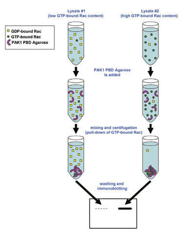
RhoA/Rac1/Cdc42 Combo Activation Assay Kit (ab211168) utilizes Rhotekin RBD and PAK1 PBD Agarose beads to selectively isolate and pull-down the active form of Rho/Rac/Cdc42 from purified samples or endogenous lysates. Subsequently, the precipitated GTP-Rho or Rac or Cdc42 is detected by western blot analysis using an anti-RhoA or Rac1 or Cdc42 antibody.The product provides enough reagents to perform 3 x 10 assays: 10 assays for each of the small GTPases (RhoA, Rac1 and Cdc42) respectively.Features: 1) non radioactive assay format; 2) fast results: 1 hour assay plus electrophoresis/blotting time; 3) includes RhoA, Rac1 and Cdc42 positive control; 4) pink colored agarose beads for easy identification during washing and aspiration steps.
- Rhoa Activation Assay Biochem Kit For Sale
- Rhoa Activation Assay Biochem Kit List
- Rhoa Activation Assay Biochem Kits
Notes. Small GTP-binding proteins (or GTPases) are a family of proteins that serve as molecular regulators in signaling transduction pathways.RhoA, Rac1 and Cdc42 are all 21 kDa proteins that belong to the family of Rho GTPases. They regulate a variety of biological response pathways that include cell motility, cell division, gene transcription, and cell transformation.
Project pat net worth. Like other small GTPases, RhoA, Rac1 and Cdc42 regulate molecular events by cycling between an inactive GDP-bound form and an active GTP-bound form. In its active (GTP-bound) state, RhoA binds specifically to the Rho-binding domain (RBD) of Rhotekin, and Rac1 or Cdc42 binds specifically to the p21-binding domain (PBD) of p21activated protein kinase (PAK) to control downstream signaling cascades.
Product Uses Include. Rho signaling pathway studies. Rho activation assays with primary cells.
Studies of Rho activators and inactivators. Rho activation assays with limited material. High throughput screens for Rho activationIntroductionThe G-LISA Rho activation assays are ELISA based Rho activation assays with which you can measure Rho activity in cells in less than 3 h.
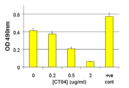
BK124 is very sensitive and has excellent accuracy between duplicate samples. For a more detailed introduction on G-LISA assays and a listing of other available G-LISA kits, see our main. The BK124 Rho activation assay kit measures the level of GTP-loaded RhoA only in cells. The level of activation is measured with absorbance set at 490nm.
For a kit to measure RhoA activation with luminescence detection, see Cat. #.See on our G-LISA page for more details.Kit contentsThe kit contains sufficient reagents to perform 96 RhoA activation assays.
Since the Rho-GTP affinity wells are supplied as strips and the strips can be broken into smaller pieces, each kit can be used for anywhere from one to multiple assays. The following components are included in the kit:.
Rho-GTP affinity wells (12 strips of 8 wells). Lysis buffer. Binding buffer. Antigen presenting buffer. Wash buffer. Antibody dilution buffer.
Anti-RhoA antibody. HRP-labeled secondary antibody. Positive control RhoA protein. Protease inhibitor cocktail (Cat.

# ). Absorbance detection reagents. Precision Red™ Advanced protein assay reagent (Cat. # ).
Manual with detailed protocols and extensive troubleshooting guide. Rho activity measured in Swiss 3T3 cells treated with the Cell Permeable Rho Inhibitor using the RhoA G-LISA Activation Assay (Cat.# BK124). Serum starved Swiss 3T3 fibroblasts were untreated (no CT04) or treated with 0.20, 0.50 and 2.0 µg/ml of for 4h in serum free medium at 37°C, then activated with 100µg/ml calpeptin for 10min. Cells were then lysed and RhoA activity was measured by the RhoA G-LISA Activation Assay (Cat.# BK124). Note: At 2.0 µg/ml CT04 for 4h results in almost complete (90%) inhibition of RhoA activity.Associated Products:Total RhoA ELISA (Cat. # )Anti-RhoA monoclonal antibody (Cat.# )Rho Inhibitor I (Cat.# )Rho Activator II (Cat.# )Rho Pathway Inhibitor I - Rho Kinase (ROCK) inhibitor Y-27632 (Cat. Herr et al., 2014.
Tetraspanin CD9 regulates cell contraction and actin arrangement via RhoA in human vascular smooth muscle cells. 9:e106999.Kalia et al., 2013. Japanese encephalitis virus infects neuronal cells through a clathrin-independent endocytic mechanism.
87, 148-162.Kanazawa et al., 2013. The Rho-kinase inhibitor fasudil restores normal motor nerve conduction velocity in diabetic rats by assuring the proper localization of adhesion-related molecules in myelinating Schwann cells. And Addison, 2012.
RhoB controls endothelial cell morphogenesis in part via negative regulation of RhoA. Vascular Cell. V 4, p 1.Yang and Kim, 2012.
The RhoA-ROCK-PTEN pathway as a molecular switch for anchorage dependent cell behavior. V 33, pp 2902-2915.Garrido-Gomez et al., 2012. Annexin A2 is critical for embryo adhesiveness to the human endometrium by RhoA activation through F-actin regulation. Doi: 10.1096/fj.12-204008.Greco et al., 2012. Chemotactic effect of prorenin on human aortic smooth muscle cells: a novel function of the (pro)renin receptor.
Cardiovasc Res. Doi: 10.1093/cvr/cvs204.Chen et al., 2012. Inhibition of tumor cell growth, proliferation and migration by X-387, a novel active-site inhibitor of mTOR. V 83, pp 1183-1194.Zhou et al., 2012. HSV-mediated gene transfer of C3 transferase inhibits Rho to promote axonal regeneration.
Et al., 2012. Protease-activated receptor 1 (PAR1) coupling to Gq/11 but not to Gi/o or G12/13 is mediated by discrete amino acids within the receptor second intracellular loop. Cellular Signalling. V 24, pp 1351-1360.Ramseyer et al., 2012.
Tumor Necrosis Factor α Decreases Nitric Oxide Synthase Type 3 Expression Primarily via Rho/Rho Kinase in the Thick Ascending Limb. V 59, pp 1145-1150.Dhaliwal et al., 2012.
Cellular cytoskeleton dynamics modulates non-viral gene delivery through RhoGTPases. V 7, e35046.Jin et al. Increased SRF Transcriptional Activity is a Novel Signature of Insulin Resistance in Humans and Mice. J Clin Invest.Ganguly et al. Adiponectin Increases LPL Activity via RhoA/ROCK-Mediated Actin Remodelling in Adult Rat Cardiomyocytes.
Rhoa Activation Assay Biochem Kit For Sale
Endocrinology 152,247.Rapier et al., 2010. Cancer Cell Int. 10, 24Nini L, Dagnino L.
Accurate and reproducible measurements of RhoA activation in small samples of primary cells. Anal Biochem 398,135-7.Yang et al. Fluoride induces vascular contraction through activation of RhoA/Rho kinase pathway in isolated rat aortas. Environmental Toxicology and Pharmacology 29,290-296.Musso et al. Relevance of the mevalonate biosynthetic pathway in the regulation of bone marrow mesenchymal stromal cell-mediated effects on T cell proliferation and B cell survival. Haematologica DOI: 10.3324/haematol.2010.031633.Lichtenstein et al. Secretase-Independent and RhoGTPase/PAK/ERK-Dependent Regulation of Cytoskeleton Dynamics in Astrocytes by NSAIDs and Derivatives.
J Alz Dis 22,1135.Ridgway et al. Modulation of GEF-H1 Induced Signaling by Heparanase in Brain Metastatic Melanoma Cells. J Cellular Biochemistry 111,1299-1309.Fang et al. Allogeneic Human Mesenchymal Stem Cells Restore Epithelial Protein Permeability in Cultured Human Alveolar Type II Cells by Secretion of Angiopoietin-1. J Biol Chem 285,2.Romero et al.
Chronic Ethanol Exposure Alters the Levels, Assembly, and Cellular Organization of the Actin Cytoskeleton and Microtubules in Hippocampal Neurons in Primary Culture. 118,602-612.Rapier et al. The extracellular matrix microtopography drives critical changes in cellular motility and Rho A activity in colon cancer cells.

Cancer Cell International 10,24.Hammar et al. Role of the Rho-ROCK (Rho-Associated Kinase) Signaling Pathway in the Regulation of Pancreatic β-Cell Function. Endocrinology 150,2072-2079.Chastre et al. TRIP6, a novel molecular partner of the MAGI-1 scaffolding molecule, promotes invasiveness.
FASEB Journal 23,916–928.Ramirez et al., 2008. 180, 1854Sequeira et al. Rho GTPases in PC-3 prostate cancer cell morphology, invasion and tumor cell diapedesis. Clinical and Experimental Metastatis 25,569-579.Moore et al.
Rho inhibition recruits DCC to the neuronal plasma membrane and enhances axon chemoattraction to netrin 1. Development 135,2855-2864.Kinoshita et al.
Apical Accumulation of Rho in the Neural Plate Is Important for Neural Plate Cell Shape Change and Neural Tube Formation. Molecular Biology of the Cell 19,2289-2299.Seifert et al. Differential activation of Rac1 and RhoA in neuroblastoma cell fractions. Neurosci Lett 450,176-180.Korobova and Svitkina (2008). Arp2/3 Complex Is Important for Filopodia Formation, Growth Cone Motility, and Neuritogenesis in Neuronal Cells. 19,1561-1574.Mercer and Helenius (2008).
Vaccinia Virus Uses Macropinocytosis and Apoptotic Mimicry to Enter Host Cells. Science 320,531.Keely et al., 2007. Methods Enzymol.
V 426, p 27.Scott et al., 2007. J Invest Dermatol. V 127, p 668.Schreibelt et al. Reactive oxygen species alter brain endothelial tight junction dynamics via RhoA, PI3 kinase, and PKB signaling. FASEB Journal 21,3666-3676.Tanaka et al. Neural Expression of G Protein-coupled Receptors GPR3, GPR6, and GPR12 Up-regulates Cyclic AMP Levels and Promotes Neurite Outgrowth.
Chem 282,5.Bradley et al., 2006. Mol Biol Cell. Question 1: Can I detect isoforms other than RhoA, Rac1 or RalA with these G-LISA activation assays?Answer 1: Yes, the RhoA G-LISA (Cat. # BK124), Rac1 G-LISA (Cat. # BK128) and RalA G-LISA (Cat.
# BK129) can be used to detect RhoB or RhoC, Rac 2 or Rac3 or RalB, respectively. The capture proteins that the wells have been coated with bind all of the isoforms of the respective GTPase. The specificity of signal is conferred by the specificity of the monoclonal primary antibody utilized. Use of an isoform-specific monoclonal antibody allows detection of other Rho family isoforms. Please see this citation for an example of this modified procedure (Hall et al., 2008.
Type I Collagen Receptor (α 2β 1) Signaling Promotes Prostate Cancer Invasion through RhoC GTPase. 10, 797–803).Basically the researcher would test their specific monoclonal antibody in a western blot first to prove specificity to the alternative isoform of interest. For example, load RhoA and C for negative controls when testing a RhoB monoclonal antibody. Then the researcher would use 1:50, 1:200 and 1:500 dilutions of their monoclonal antibody on duplicate cell extracts of activated and control state samples. The researcher would then choose the dilution of monoclonal antibody which gave them the highest ratio of activated:control state.A simple activated/control state pair of extracts can be made by growing cells to 50% confluence in serum containing media, washing twice with PBS, preparing lysate and aliquoting and freezing samples in liquid nitrogen. With one aliquot, defrost and let stand at room temperature for 60 min to degrade the activated signal to a low basal signal, which will be the control state.
Rhoa Activation Assay Biochem Kit List
The untreated sample (2nd aliquot) will be considered “activated” which most serum grown cells are.Question 2: How many cell culture plates can I process at one time during the lysis step?Answer 2: We recommend that from the point at you add lysis buffer to the plate on ice to aliquoting and snap-freezing the lysate samples in liquid nitrogen, no more than 10 min are allowed to elapse. After 10 min on ice, we find that GTP bound to GTPases (activated GTPases) undergoes rapid hydrolysis. Rapid processing at 4°C is essential for accurate and reproducible results. The following guidelines are useful for rapid lysis of cells.Washinga.
Retrieve culture dish from incubator, immediately aspirate out all of the media and place firmly on ice.b. Immediately rinse cells with an appropriate volume of ice cold PBS (for Cdc42 activation, skip this step and simply aspirate the media) to remove serum proteins.c. Aspirate off all residual PBS buffer. This is essential so that the Lysis Buffer is not diluted. Correct aspiration requires that the culture dish is placed at a steep angle on ice for 1 min to allow excess PBS to collect in the vessel for complete removal.
As noted, the time period between cell lysis and addition of lysates to the wells is critically important. Take the following precautions:1. Work quickly.2. Keeping solutions and lysates embedded in ice so that the temperature is below 4°C. This helps to minimize changes in signal over time.3. We strongly recommend that cell lysates be immediately frozen after harvest and clarification.

Rhoa Activation Assay Biochem Kits
A sample of at least 20 μl should be kept on ice for protein concentration measurement. The lysates must be snap frozen in liquid nitrogen and stored at -70°C. Lysates should be stored at -70°C for no longer than 30 days.4. Thawing of cell lysates prior to use in the G-LISA assay should be in a room temperature water bath, followed by rapid transfer to ice and immediate use in the assay.If you have any questions concerning this product, please contact our Technical Service department at.